Image Challenge
To promote critical thinking as well as friendly rivalry, the image challenge was implemented. By distributing images with a clinical vignette, residents and faculty test their medical knowledge by answering questions of diagnoses and management.
Challenge 1
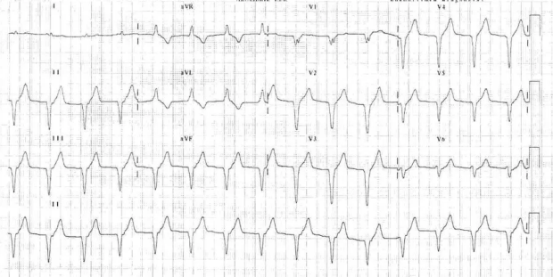
Vignette: A nurse calls you regarding an abnormal rhythm on a 62 yo M who recently had a STEMI. You obtain the EKG noted here. What is the underlying rhythm (1 pt) and what should you do about it? (1pt)
Flip panel for explaination
Solution
1) Rhythm: AIVR (accelerated idioventricular rhythm)
2) Management: No specific management needed.
Further Discussion
The rhythm seen above is AIVR, which is accelerated idioventricular rhythm, a regular monomorphic ventricular rhythm usually with a rate in the 60s-100s. This is a common rhythm that occurs after myocardial reperfusion (as in the case of the STEMI). It occurs when the rate of an ectopic ventricular pacemaker exceeds that of the sinus node. You may see occasional fusion and capture beats. Because this rhythm is at a well tolerated rate and usually self limiting, nothing beyond usual management of the underlying issue is needed. In fact, antiarrhythmics may precipitate worse arrhythmias.
Challenge 2
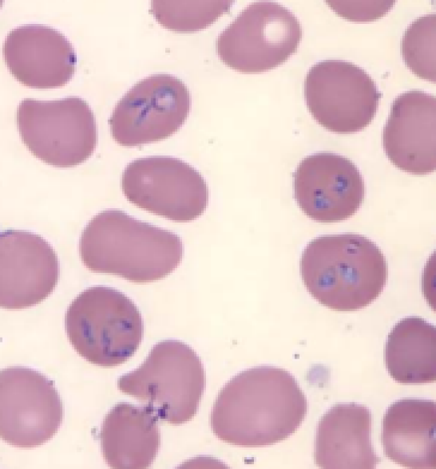
Vignette: A 64 year-old previously healthy woman presents to the ED after being found down in her sister’s home. She is visiting the Bay Area for a wedding but has been feeling ill for roughly the last week with mild headache, fatigue, myalgias and daily temperatures to 101-102F. The day prior to presentation she started reporting abdominal pain with nausea. Today she was found by her sister unresponsive on the floor of her home; her sister noted she appeared pale and called EMS. In the ED she was found to be hypotensive and anemic. FAST exam revealed free fluid and she was taken urgently to the OR where she underwent emergent splenectomy for a spontaneous splenic rupture. Peripheral smear was done on her initial CBC as noted. What infection does this patient have (1 point) and what is the vector of transmission (1 point)?
Flip panel for explaination
Solution and Explanation
This woman had a babesia infection (1 point), which is transmitted most commonly by the Ixodes scapularis tick (1 point). The transmission of this infection occurs most commonly in the upper Midwest and northeast of the United States, primarily in the summer months when the ticks are still in the nymph stage. Transmission can rarely occur with blood transfusion and solid organ transplant.
On peripheral smear, babesia is characterized by intracellular parasites. While the Maltese crosses are pathognomonic for the infection, they are not always seen.
Severity of Infection:
- Mild Infection (most common): relatively non-specific: fever, myalgias, malaise, chills
- Severe infections: heart failure, renal failure, ARDS and spontaneous splenic rupture. GI symptoms (N/V/D) are predictive of severe infection. Risk factors for severe infection are related to older age, asplenism or other immunocompromised state.
- This woman clearly met severe infection. Can reference case report at PMID 26123434 for further details.

Challenge 3
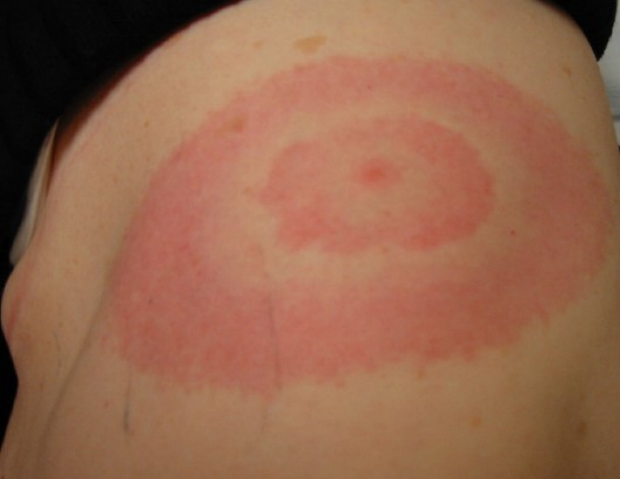
Vignette: A 21 y/o M presents with fevers, abdominal pain, and the following rash. He recently went on a post college celebratory trip with his friends abroad. What is the underlying diagnosis?
Flip panel for explaination
Solution: Salmonella enterica/typhi (Enteric typhoid fever)
Discussion: This patient had enteric fever (not to be confused with epidemic typhus which is transmitted by the body lice) and the rash is a classic "rose spot rash", which is pathognomonic
for this disease. Enteric fever is a very prevalent infectious disease with fecal-oral transmission worldwide. The main manifestations to look for in a patient with this disease is a stepwise presentation. During the first week of presentation, patients will have fevers and "pulse-temperature" dissociation in which relative bradycardia can be observed. The second week is typically when patients develop abdominal symptoms including diarrhea and/or constipation as well as the rash seen above. If illness is severe, patients can develop shock, perforation, peritonitis and hemorrhage in the third week. Diagnosis is made via cultures most commonly of the blood but stool and urine can also be cultured. In fact, bone marrow cultures are the most sensitive for finding the organism. Usually, empiric 3rd generation cephalosporins can be used until culture results return but there has been rising antimicrobial resistance and XDR Salmonella enterica has been found in Pakistan.
Challenge 4
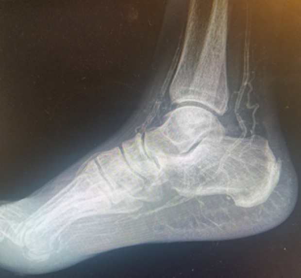
Vignette: An elderly woman with ESRD presents with pain in all her limbs. You obtain this xray of her foot. What is the diagnosis?
Flip panel for explaination
Solution and Discussion
The xray demonstrates vascular calcification. This woman has calcific uremic arteriolopathy, commonly known as calciphylaxis.
This typically happens in ESRD patients – these patients have deposition of calcium salts in cutaneous blood vessels, which eventually lead to ischemic tissue necrosis (and pain).
Challenge 5
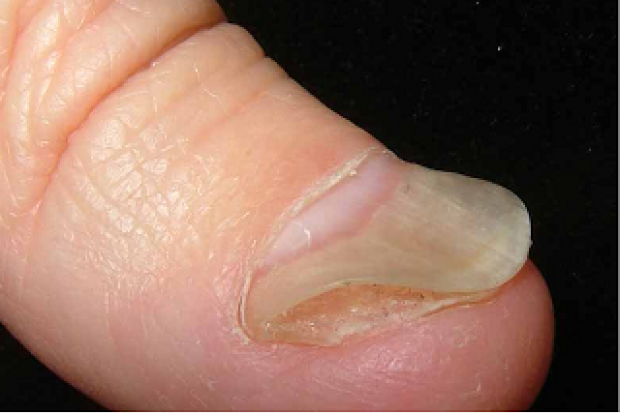
Vignette: A 34 yo F presents to you with fatigue. On physical exam, you note the finding in the image. What's the underlying etiology?
Flip panel for explaination
Solution
Koilonychia (spoon nail) likely secondary to iron deficiency anemia
Discussion
This patient has koilonychia, which is the thinning of the nails causing a flattened and often concave up shape. Most commonly, this is associated with iron deficiency anemia though can be inherited or associated with trauma and chemical exposure.
Challenge 6
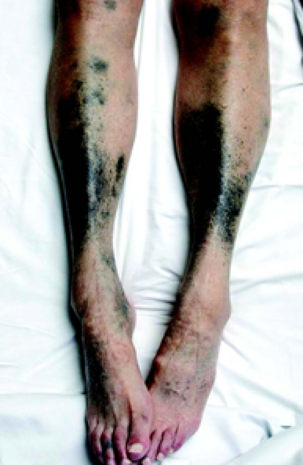
Vignette: A 65-year-old woman with history of prior endocarditis s/p aortic valve replacement with porcine valve complicated by recurrent episodes of endocarditis presents with the above painless skin findings. Since treatment of her last episode of endocarditis she has been feeling well with no constitutional symptoms and no pain in her legs. Which medication may she be taking that leads to this leg discoloration?
Flip panel for explaination
Solution: Minocycline
Discussion:
For reasons that are not understood, when minocycline is oxidized it turns black, which can lead to discoloration of teeth, conjunctivae, skin and nails. This is more likely to occur if patients are on concurrent amitriptyline or estrogen. Treatment is discontinuation of the minocycline, but it can still take months to years to resolve.
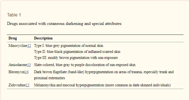
Table 1 from attached study (PMID 23754872)
Solution: Minocycline
Discussion:
For reasons that are not understood, when minocycline is oxidized it turns black, which can lead to discoloration of teeth, conjunctivae, skin and nails. This is more likely to occur if patients are on concurrent amitriptyline or estrogen. Treatment is discontinuation of the minocycline, but it can still take months to years to resolve.
Table 1 from attached study (PMID 23754872)
Challenge 7a
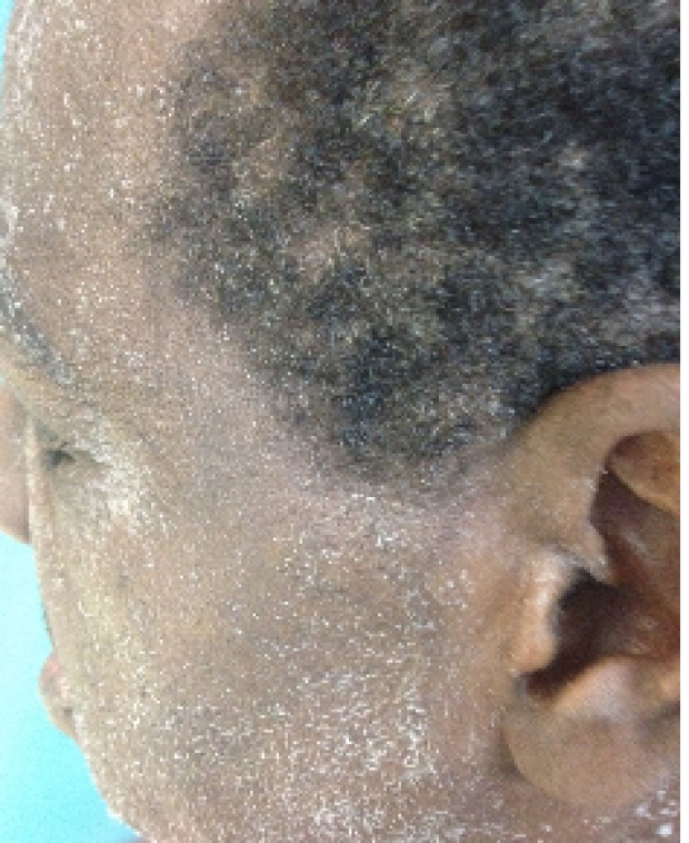
Vignette: A 52 yo M presents with AMS and nausea. On exam, you notice findings in the image. What's the finding on exam and the cause of the patient's presentation?
Flip panel for explaination
Solution: Uremic Frost
Discussion: In patients with CKD and azotemia, the high levels of urea in the body is secreted as part of sweat. After the water evaporates, the remaining urea crystallizes and you end up with uremic frost. This typically happens in patients with BUN>200 and is becoming rarer.
Challenge 7b
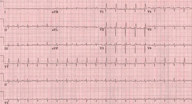
Vignette: A 48 yo M presents to you for a work physical. He is otherwise asymptomatic. You obtain an EKG, shown here. What's the cause of the patient's EKG findings?
Flip panel for explaination
Solution: transplanted heart
Further discussion: In this EKG you see that it is sinus rhythm but with anomaly of two distinct sets of p waves, only one of which is actually being conducted. This occurs in the setting of a transplanted heart where there is a remnant of the recipient's atria/sinus node.
Challenge 8
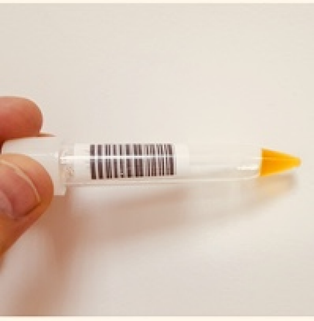
Vignette: A 62 yo F who presents with altered mental status and lower extremity weakness. You perform a lumbar puncture but then the CSF ends up looking like the image findings noted. What is the phenomenon called and what is its etiology?
Flip panel for explaination
Solution: Nonne-Froin's syndrome or Froin's sign
Further explanation: In the above picture, you see that the CSF is coagulated and not flowing with gravity as well as has the distinctive yellow color of xanthochromia. This occurs when there is a high level of protein in the CSF, commonly caused by tumor/mass occlusion. It is an uncommon finding. Many of you gave the answer of xanthochromia. However, that is only a partial solution and the coagulation of the CSF seals the diagnosis as Nonne-Froin's syndrome.
Challenge 9
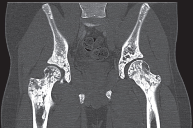
Vignette: A 19-year-old man had a CT scan performed, which incidentally showed these bone findings (patient asymptomatic). Diagnosis?
Flip panel for explaination
Solution: osteopoikilosis or “spotted bone disease.”
Discussion: This is an autosomal dominant disorder that is characterized by the above benign sclerotic bone lesions. Bone lesions are painless (differentiates from mets) and distribution is typically symmetric (also differentiates from mets) in the pelvis, hands, feet and ends of long bones.
Challenge 10
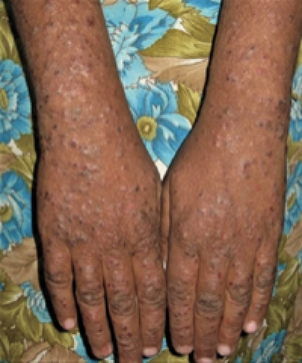
Vignette: A 27 y/o previously healthy woman presents with the pruritic lesions noted here. What is the underlying infection?
Flip panel for explaination
Solution: This is called pruritic papular eruption (PPE) and her underlying infection is HIV (most often seen in patients with low CD4 counts).
Discussion: PPE is characterized by bilateral and symmetric distribution of very pruritic papules on the trunk and extremities (not entirely on sun exposed areas). PCT (porphyria cutanea tarda) can look very similar and key in distinguishing the two would be the distribution of the rash. (photosensitive areas for PCT)
Challenge 11
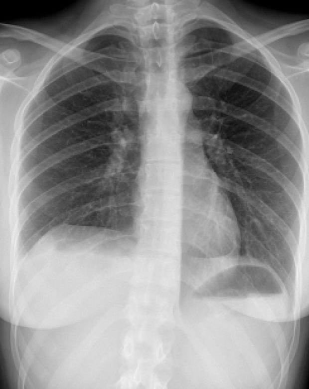
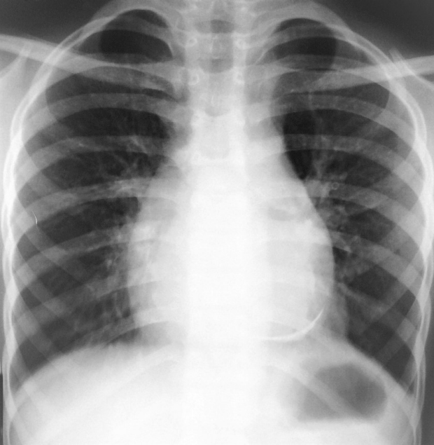
Vignette: A 53 yo F with prior hx only significant for asthma and IBS presenting with SOB. Her CXR is shown here. What's the imaging finding (1pt) and the underlying diagnosis? (1pt)
Flip panel for explaination
Solution: Westermark sign, indicating acute pulmonary embolism
Discussion: The Westermark sign is found on plain chest xrays where you see an area of peripheral hyperlucency secondary to an area of decreased blood flow from the embolus. In studies, this sign is present in 2-10% of patients with confirmed PEs. Sensitivity of this sign is low (~10%) but specificity is high (~90%).
Challenge 12
Vignette: A 24 y/o woman returns to Palo Alto after spending the holidays with family in the midwest. She has developed this rash. What underlying organism causes this rash?
Flip panel for explaination
Solution and Discussion:
This rash is called erythema migrans and is seen in roughly 80% of patients who develop Lyme Disease. The underlying organism was Borrelia burgdoferi. Notably, this infection is transmitted via the Ixodes tick, and patients may have co-infection with babesiosis (Ixodes tick transmits both babesiosis and Lyme Disease).
Challenge 13
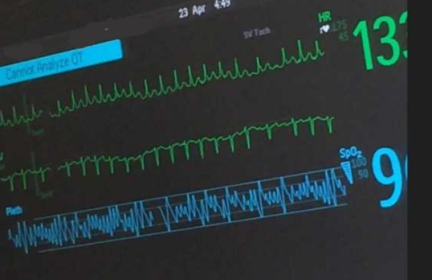
Vignette: You are working nights and you suddenly get a page for an rapid response team for a patient. It is for a patient on med 10 who has shortness of breath, tachycardia to the 130s and borderline hypotension with BP 96/74. He looks clearly in distress. His monitor is noted here. What is going on?
Flip panel for explaination
Solution: Cardiac tamponade
Discussion: The patient developed atrial fibrillation with RVR as noted on the telemetry monitor. The pulse ox demonstrates evidence of varying of the waveform with respiration suggestive of a variant of electrical alternans. Often, especially in patients with arterial lines, the alterations in the waveform will alert astute clinicians of the possibility of tamponade without formal measurement of a pulsus. This patient ultimately received an urgent pericardiocentesis.
Challenge 14
Vignette: A 41 y/o previously healthy gentleman presents to the ED with 3 days of chest pain. This is his CXR. Diagnosis?
Flip panel for explaination
Solution and Discussion:
His CXR demonstrated evidence of calcifications in his pericardium, making the most likely diagnosis pericarditis. Pericardial effusion with water bottle sign was also accepted as a correct answer.