Musculoskeletal Ultrasound Clinic Is a BOON to Patient Care, Education, and Research
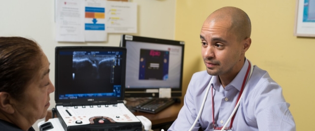
Rob Fairchild, MD, uses ultrasound for many diagnostic and treatment purposes, including evaluating inflammatory arthritis.
As a fellow in immunology and rheumatology, Rob Fairchild, MD, noticed something lacking in the care of rheumatology patients, and he set out to change that.
“The use of ultrasound by rheumatologists is more common in Europe than in the United States,” Fairchild observed. He was intrigued because ultrasound is a relatively easy tool that can be performed quickly in the clinic, and it’s an effective means for viewing soft tissue and other structures that can help rheumatologists with diagnosis and treatment.
“I did some training on ultrasound during my first year of fellowship, and that led me to devote one of my fellowship electives to starting a musculoskeletal ultrasound clinic dedicated to rheumatology evaluations and interventions,” he says.
Now, as the newest full-time member of the immunology and rheumatology faculty, Fairchild is seeing that the clinic continues not only for the benefit of patients, but also for the education of other trainees.
In fact, the American College of Rheumatology is moving toward incorporating ultrasound as part of rheumatology training, so Fairchild will be building that training into the fellowship curriculum.
The Craft So Long to Learn
The rheumatologist admits that ultrasound is very complicated and takes a long time to master. It requires learning separate views for each of the joints, and there are a lot of structures to know.
But ultrasound has long been an effective and accepted modality among many specialties, so what makes the Rheumatology Ultrasound Clinic distinct from other musculoskeletal ultrasound clinics?
“There’s actually a really big distinction. First and foremost, I’m a rheumatologist/immunologist. While most specialties use musculoskeletal ultrasound for soft tissue ailments like tendonitis, bursitis, and other joint abnormalities, rheumatologists are also trained to evaluate and manage conditions specific to our field, such as inflammatory arthritis or gout. So, we are often looking for very different things than other ultrasonographers.”
While Fairchild heads the clinic, two other attending rheumatologists—Jison Hong, MD, and Janice Lin, MD—also perform several procedures.
Evaluation and Treatment
Ultrasound helps Fairchild, Hong, and Lin when they are on the lookout for unusual disease manifestations like glandular disease in Sjogren’s syndrome, a debilitating condition that causes the eyes, mouth, or other parts of the body to dry out. It’s also useful in diagnosing polymyalgia rheumatica, an inflammatory disorder that causes muscle pain and stiffness, especially in the shoulders and hips. And ultrasound is a great aid in looking at temporal arteries to spot giant cell arteritis, which, if left untreated, can lead to blindness.
Two of the most frequent referrals the clinic receives are inflammatory arthritis evaluations and interphalangeal joint injections of the hands.
In one recent case Fairchild was asked to evaluate whether there was evidence of an underlying inflammatory arthritis in a patient, as diagnosed by the patient’s previous rheumatologist.
According to Fairchild, “The patient had been on significant immunosuppression with a combination of steroids, methotrexate, and weekly TNF-alpha inhibitor injections, which all have the potential for serious side effects, require frequent clinical and laboratory monitoring, and are expensive. Our clinic’s ultrasound evaluation of the hands showed no synovial hypertrophy, synovitis, joint effusion, or erosions, which are the hallmarks of rheumatoid arthritis. Using this additional information coupled with the patient’s history and clinical evaluation, the referring provider at Stanford felt confident that the patient’s immunosuppression was not warranted and began to wean the patient off the medications.”
Another recent referral was for intra-articular injections for severe inflammatory/erosive osteoarthritis. “This aggressive, debilitating disease causes severe damage to the distal joints of the fingers with bony proliferation coupled with inflammation, pain, and dysfunction,” he explains.
“One way to reduce swelling, pain, and inflammation in these joints is through steroid injection. However, these joints are very small, and needle injection can be quite painful and technically challenging because of the bony mass surrounding the joint, making needle guidance difficult. When referred these patients, I use ultrasound to accurately guide the needle into the joint in one pass, greatly improving procedure tolerability and accurate steroid placement. As a testament to the efficacy and tolerability of these ultrasound guided procedures, I frequently have patients request repeat visits for additional therapeutic intervention once the steroid has worn off,” he says.
'Old' and 'New School' Practitioners
Not everyone is convinced of the value of ultrasound.
Many rheumatologists are familiar and comfortable with “classic” examination techniques like feeling a patient’s joints for warmth, swelling, and tenderness to make an excellent diagnosis. “That’s very different from the ‘new school’ practitioners who can pull out an ultrasound and combine it with a clinical exam to give even greater accuracy. A lot of ‘old school’ rheumatologists would balk at that, but studies have shown that ultrasound is superior in finding active disease, particularly when the disease is mild, where it can be missed with a clinical exam alone,” Fairchild notes.
His interest in ultrasound is convincing other, more established rheumatologists that this technique is important for everyone to know and incorporate into their practice. In fact, some providers who may not have appreciated the value of ultrasound initially are now warming up to it.
Plans for Research
Several areas of research fit into Fairchild’s plans for the clinic. One has to do with how patients perceive their disease when they see it by ultrasound.
“I can tell patients that their disease is really active as a means of encouraging them to take a very serious medication, but that’s quite different from putting an ultrasound on them, pointing to the inflamed area and showing them how the joint is abnormal or damaged. They have an immediate response to that,” he says when explaining his desire to develop a research project in that area.
Another research interest involves scleroderma patients, who can be very sick with soft tissue and skin manifestations. There’s been a lot in the literature recently that has looked at ultrasound and how it can be used to assess disease, severity, the kind of disease that the patient actually has, and how it can help with treatments. Fairchild is pursuing a project in that realm with Lorinda Chung, MD, MPH, who runs Stanford’s Autoimmune Skin Disease Clinic in Redwood City with David Fiorentino, MD, PhD.
Training Tomorrow's Ultrasonographers
Resident and fellow training is another facet of the ultrasound clinic.
“Coupled with their training in the clinic, we also do training at the bedside as part of Stanford 25,” says Fairchild. “Last year Dr. Hong and I did several musculoskeletal ultrasound teaching sessions for the residents in the hospital—hands-on things to show them how to look for knee effusions and other simple things that would be useful on the floor. I want to try to expand that as much as possible in the future.”